
Current Issue: July 2026
Editor's Choice
This month's highlight articles
Low-dose rectal indomethacin to prevent post-ERCP pancreatitis: results of a large Chinese multicenter randomized noninferiority trial
Graphical Abstract
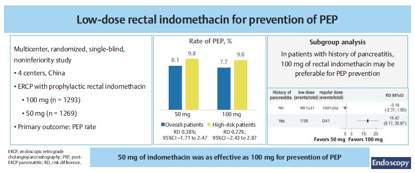
Table of Contents
Author commentary
E-Videos
Letter to the editor
Letter to the editor
Letter to the editor
Original article
Original article
Original article
Original article
Original article
Original article
Position Statement
Original article


Official organ of ESGE
Journal Impact Factor: 11.8
In this issue
Newsletter
Don't want to miss important information?
Sign up for your monthly Endoscopy-Newsletter: Our Editorial Team offers insight in what's new and important.